Foot Bone Chart
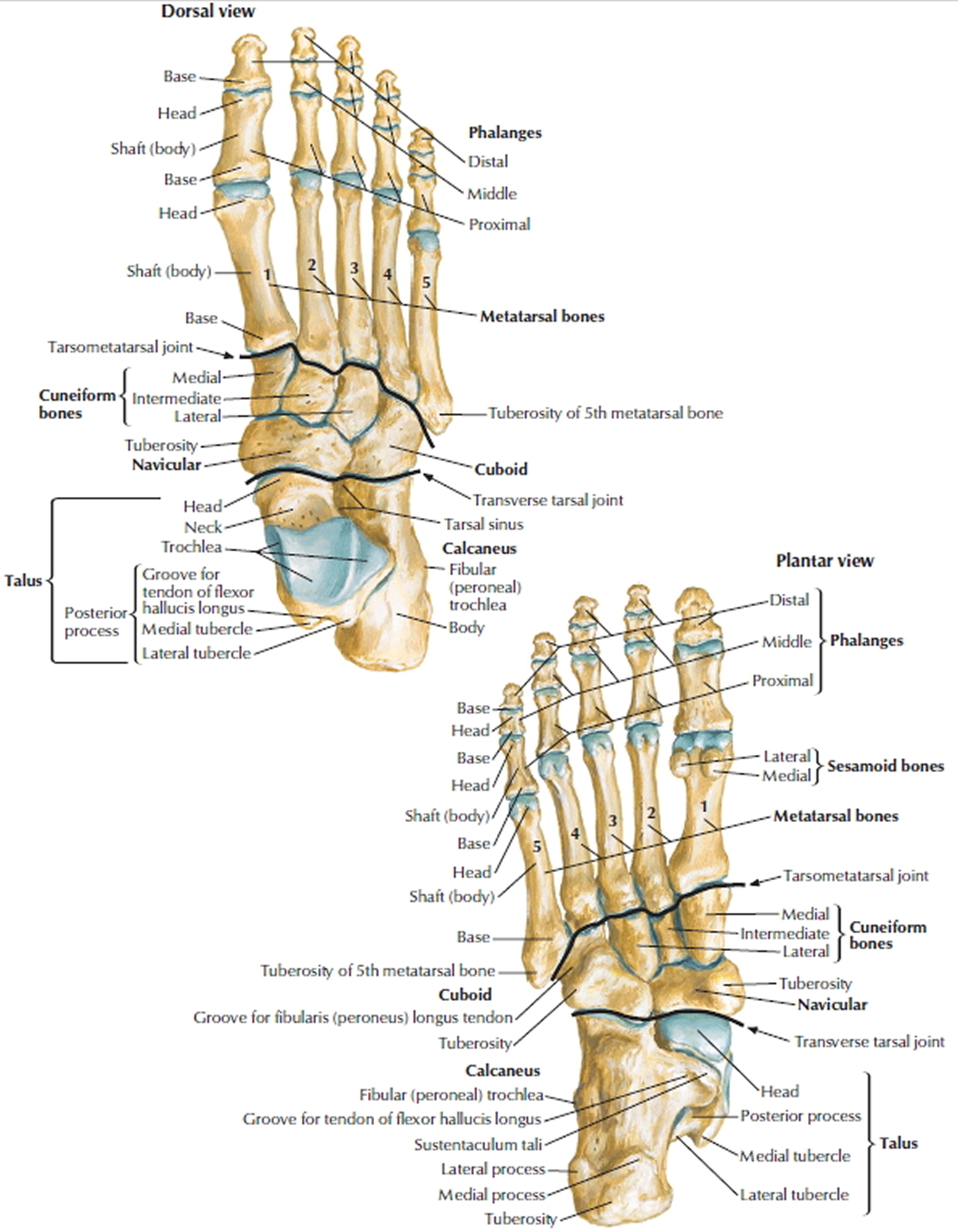
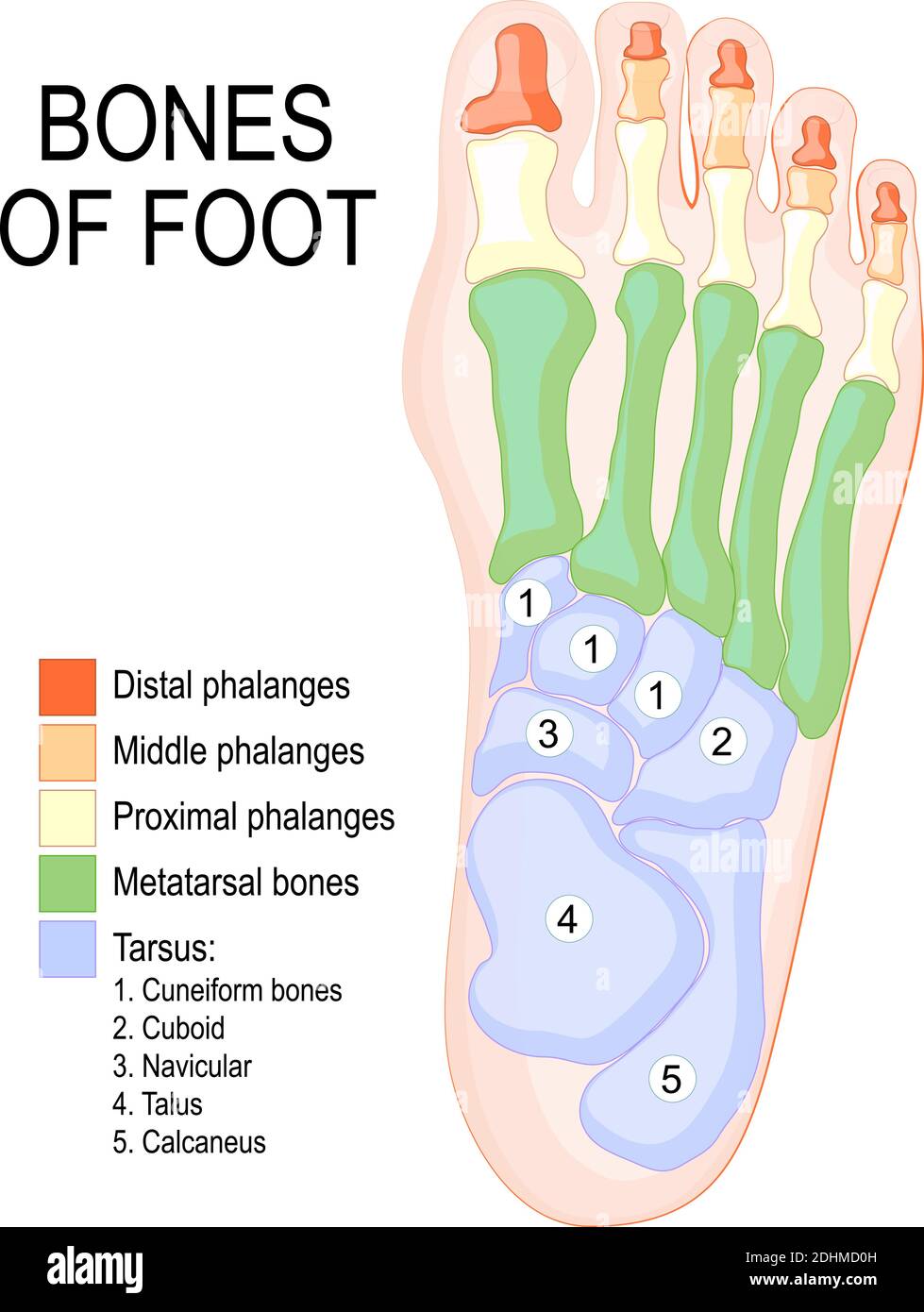
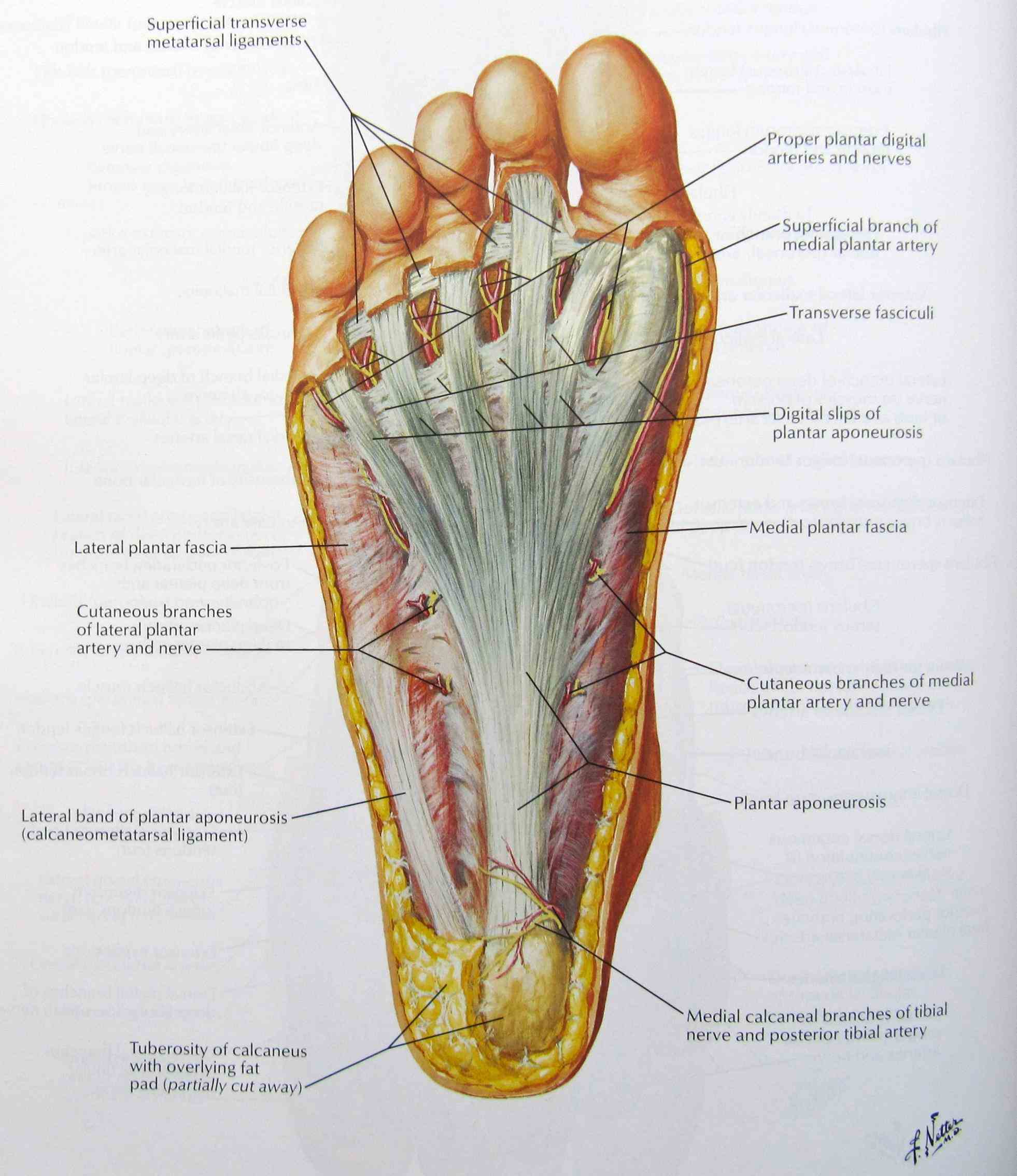
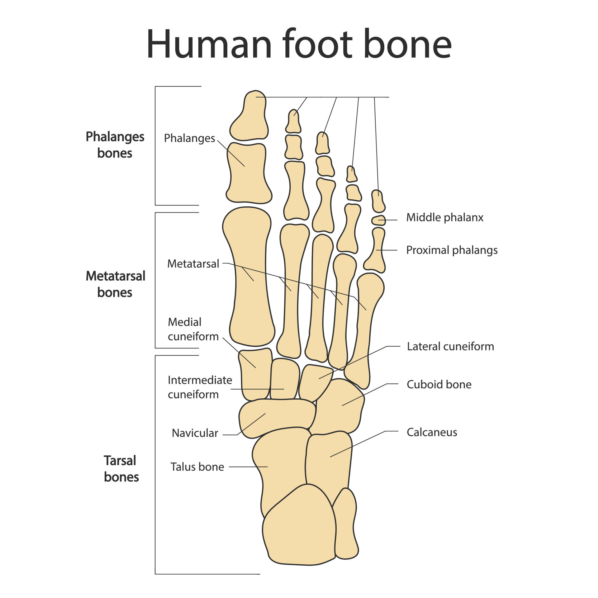
Foot Bone Chart - The top of the foot is referred to as the dorsal surface. This article will describe in detail the anatomy and function of the major bones in the foot. It functions as a rigid structure for weight bearing and it can also function as a flexible structure to conform to uneven terrain. The hindfoot, the midfoot, and the forefoot. Web there are 26 bones in the foot. Web use our anatomy tools to learn about bones, joints, ligaments, and muscles of the foot and ankle. The sole of the foot is the plantar surface. The forefoot is composed of the metatarsals, phalanges, and sesamoids. The proximal tarsal bones are the talus and calcaneus. Major (2nd most important) medial arch support. Footeducation is committed to helping educate patients about foot and ankle conditions by providing high quality, accurate, and easy to understand information. The foot begins at the lower end of the tibia and fibula, the two bones of the lower leg. It functions as a rigid structure for weight bearing and it can also function as a flexible structure to conform to uneven terrain. Major (2nd most important) medial arch support. The distal tarsals are the cuboid and three cuneiform bones (lateral, intermediate, and medial). Learn more about foot bones and foot anatomy here. There are a whole range of structures e.g. Web the bones of the foot are organized into rows named tarsal bones, metatarsal bones, and phalanges. Web use our anatomy tools to learn about bones, joints, ligaments, and muscles of the foot and ankle. The knuckles of the toes are called the metatarsophalangeal joint. Web there are 26 bones in the foot and 33 joints in the foot. These make up the toes and broad section of the feet. The sole of the foot is the plantar surface. The knuckles of the toes are called the metatarsophalangeal joint. Base of the 5th metatarsal (lateral band), plantar plate and bases of the five proximal phalanges. The sole of the foot is the plantar surface. Body weight supported by the foot is spread across the arches from the tarsal and metatarsal bones, which make contact with the ground. The bottom part of the foot is the sole. The bones that make up the forefoot are those that are last to leave the ground during walking. You. Web the foot bones account for a quarter of all the bones in our body. Web the foot can also be divided up into three regions: Footeducation is committed to helping educate patients about foot and ankle conditions by providing high quality, accurate, and easy to understand information. Web the calcaneus, or heel bone, is the largest tarsal bone and. It functions as a rigid structure for weight bearing and it can also function as a flexible structure to conform to uneven terrain. The bottom part of the foot is the sole. The hindfoot, the midfoot, and the forefoot. Footeducation is committed to helping educate patients about foot and ankle conditions by providing high quality, accurate, and easy to understand. Bones, muscles, tendons and nerves which will each give slightly different foot pain symptoms. Base of the 5th metatarsal (lateral band), plantar plate and bases of the five proximal phalanges. Body weight supported by the foot is spread across the arches from the tarsal and metatarsal bones, which make contact with the ground. The bones that make up the forefoot. Find out how the different foot bones fit together and how they are commonly injured. Web the foot and ankle form a complex system which consists of 28 bones, 33 joints, 112 ligaments, controlled by 13 extrinsic and 21 intrinsic muscles. This article will describe in detail the anatomy and function of the major bones in the foot. Web the. The forefoot, midfoot, and hindfoot. The important structures of the ankle can be divided into several categories. This complex network of structures fit and work together to bear weight, allow movement and provide a stable base for us to stand and move on. Web learn the bones of the foot in half the time with these interactive quizzes and labeling. Bones, muscles, tendons and nerves which will each give slightly different foot pain symptoms. These make up the toes and broad section of the feet. Body weight supported by the foot is spread across the arches from the tarsal and metatarsal bones, which make contact with the ground. The hindfoot, the midfoot, and the forefoot. This article will describe in. This complex network of structures fit and work together to bear weight, allow movement and provide a stable base for us to stand and move on. Web the bones of the foot are organized into the tarsal bones, metatarsal bones, and phalanges. With a pen or pencil pointed straight down, trace the outline of your foot on the paper. Footeducation. Web the foot and ankle form a complex system which consists of 28 bones, 33 joints, 112 ligaments, controlled by 13 extrinsic and 21 intrinsic muscles. Web the bones of the foot are organized into rows named tarsal bones, metatarsal bones, and phalanges. Major (2nd most important) medial arch support. Web tape a piece of paper to a hard floor,. The phalanges, which are the bones in your toes. The proximal tarsal bones are the talus and calcaneus. The bones that make up the forefoot are those that are last to leave the ground during walking. The top part of your foot above the arch is the instep. Bones, muscles, tendons and nerves which will each give slightly different foot pain symptoms. Web tape a piece of paper to a hard floor, ensuring the paper doesn’t slip. You can also sit in a chair, but make sure your feet are firmly planted on the ground. Web how the ankle works. The knuckles of the toes are called the metatarsophalangeal joint. They are complex structures with 26 bones. There are bones, joints, muscles, tendons, and ligaments in each of these sections. Web the foot can also be divided up into three regions: With a pen or pencil pointed straight down, trace the outline of your foot on the paper. Web the 26 bones of the foot consist of eight distinct types, including the tarsals, metatarsals, phalanges, cuneiforms, talus, navicular, and cuboid bones. Web this article looks at the structure of the foot — including bones, muscles, ligaments, and tendons — and some of the common conditions that affect it. Web the five bones of the midfoot comprise the navicular, cuboid, and the three cuneiforms (medial, middle, and lateral).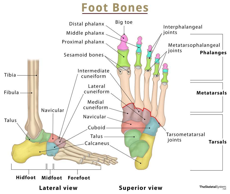
Foot Bones Names, Anatomy, Structure, & Labeled Diagrams

Bone Structure Of Foot
.jpg)
Foot Bones Labeled
Human Skeleton Skeletal System Function, Human Bones

The bones in the foot inferior view (Picture illustrated from Thieme

Foot & Ankle Bones

Anatomy of the Foot and Ankle OrthoPaedia
Bones of foot. Human Anatomy. The diagram shows the placement and names
Anatomy The Bones Of The Foot
Foot bones. Anatomy of the skeletal system of the human legs and feet
This Consists Of Five Long Bones (Metatarsal Bones) And Five Shorter Bones That Form The Base Of The Toes (Phalanges).
Web Foot And Ankle Anatomy Consists Of 33 Bones, 26 Joints And Over A Hundred Muscles, Ligaments And Tendons.
Web The Foot Bones Account For A Quarter Of All The Bones In Our Body.
Base Of The 5Th Metatarsal (Lateral Band), Plantar Plate And Bases Of The Five Proximal Phalanges.
Related Post: